Mole Mapping
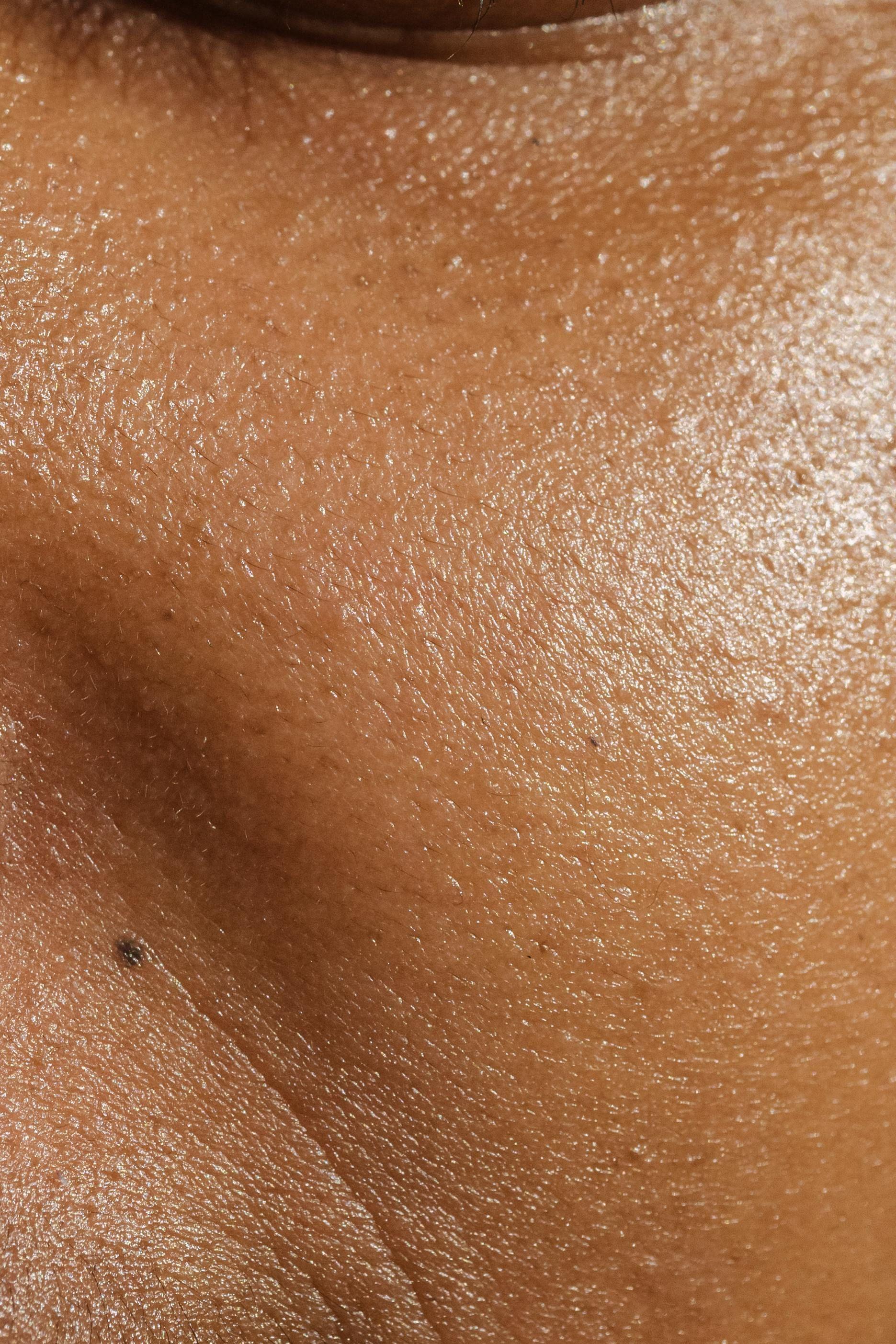
A mole or melanocytic naevus is formed of a cluster of pigment producing cells called melanocytes. Some moles may be present from birth and we call these congenital naevi.


What are moles?
A mole or melanocytic naevus is formed of a cluster of pigment producing cells called melanocytes. Some moles may be present from birth and we call these congenital naevi.
Other common moles
Junctional Naevus
These are flat moles with dark uniform pigment. The naevus cells sit at the junction of the epidermis and dermis.
Dermal Naevus
The naevus cells sit in dermis creating a raised domed mole often with a light brown or pink colour.
Compound Naevus
Naevus cells sit in dermis but also the dermo-epidermal junction creating a raised pigmented lesion
Combined Naevus
These have a mixed pattern of 2 different moles in one lesion.
Blue Naevus
The naevus cells sit deep in the dermis creating a blue tint to the pigmentation seen on the skin.
Mole Mapping
00
00
What is Mole Mapping?
What is Mole Mapping?
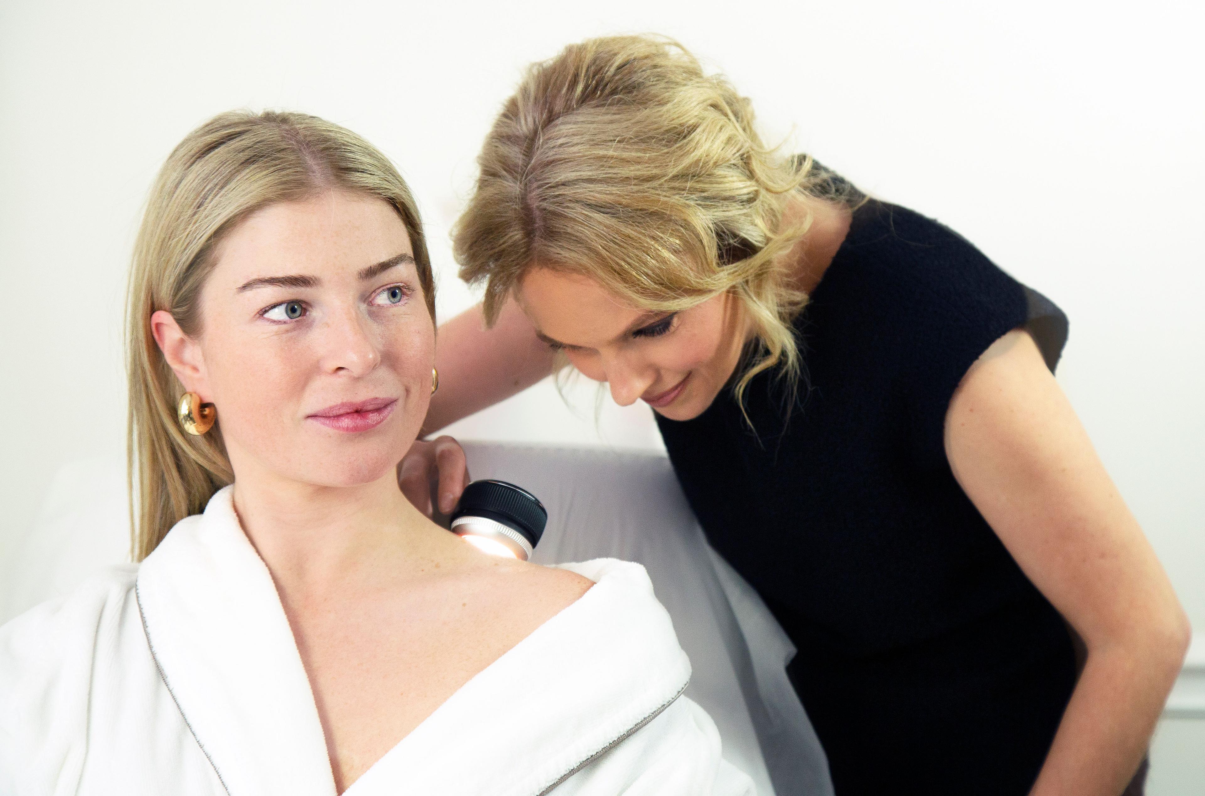
Mole mapping is a technique which catalogues or 'maps' a persons moles. The images taken by the mole mapping machine are then used as a key element in the individuals skin surveillance programme. We use the Fotofinder system and where appropriate, digital dermascopic imaging (dermascopy) to investigate any suspicious moles, changes in the skin and also new moles and lesions.

01
01
What is a Dermascope?
What is a Dermascope?
A Dermascope is a piece of equipment used to magnify your moles and other lesions to check their size, shape and pattern, and to identify any changes when regular mole mapping takes place. It uses a high quality lens at 10-14 times magnification and a lighting system which enables visualisation of sub-surface structures and patterns, which are usually not visible with the naked eye. Dermascope imaging enables trained clinicians to distinguish non-cancerous from cancerous moles and blemishes. This is most helpful in the diagnosis of skin melanoma which is the most concerning type of skin cancer.

02
02
Self Surveillance
Self Surveillance
There are differences of opinion about the recommended frequency for self surveillance of your moles. For high risk individuals, self surveillance should be undertaken at least every 1-3 months. Where there are few risk factors for melanoma a less frequent regime of self surveillance could be used.

03
03
Professional Surveillance
Professional Surveillance
To assess changes over time, skin cancer screening appointments are generally recommended every 6-12 months. Dr Catherine will advise on the time frame when it is best to schedule your mole mapping appointments according to your skin type.

FAQs
Many people worry about moles and don't know what to look out for to distinguish between normal and worrying moles. There are some commonly asked questions answered below, if you have a mole which is new, changing or causing you concern, you can book a consultation below.
Have another question?
Contact usWhy do moles become cancerous?
In around a third of cases melanoma can occur on the skin from a pre-existing mole, but it is actually more likely to present as a completely new lesion on the skin. Understanding the steps that occur within a normal mole to turn it cancerous has been critical in allowing us to develop treatments for melanoma. We know that a complex series of ‘mutations’ need to occur within the DNA of the melanocyte, making the cell behave inappropriately and evade the daily ‘immune surveillance’ that removes pre-cancerous cells from your body.
Who typically gets moles, and who is at a higher risk of developing cancerous moles
Everyone will tend to have at least 1 mole. Some people are born with them, others develop moles through childhood. Further moles that develop in adulthood tend to follow sun exposure. As a general rule adults can develop new moles until the age 40, but new pigmented ‘mole-like’ lesions appearing after this age should be reviewed by your dermatologist. Risk factors for developing cancerous moles or melanoma include your genetics, your number of moles, skin type and the way your skin responds to UV exposure (your skin type) and the level of UV you have been exposed to- particularly multiple sunburn episodes during childhood and high levels of UV exposure.
Is there anything in particular we should be looking out for and when should we seek help?
Remember that melanoma is often not sore or itchy, so if you are not looking you simply don’t notice it. I recommend patients focus on the ‘ugly duckling sign’. This is usually the first lesion your eye is drawn to when checking your skin, as it will stand out as being different from the other moles on your body. You can then focus on these lesions with a more detailed ABCD approach (Asymmetry, Boarder irregularity, colour variation and diameter >6mm). Other signs would be bleeding, crusting or a new mole appearing over the age of 40 years. If you see any of the signs listed above it is important you see your doctor or dermatologist quickly to have an assessment of the mole. Don’t delay- its natural to feel anxious but important that you have it properly assessed as soon as possible.
Is it safe to remove a mole for aesthetic reasons?
There may be moles you don’t like the look of or moles that become inflamed after rubbing on clothing. It is safe to remove these lesions for aesthetic reasons. Moles can be surgically excised –where the mole is cut out of the skin and the skin is repaired with stitches leaving a scar in its place. If a mole is raised and felt to be ‘benign’ by your dermatologist options include a ‘shave excision’ – which only removes the top section of the mole, leaving mole cells under the surface of the skin. It is important that any moles removed are histologically reviewed to confirm diagnosis.
Are there any areas we should pay particular attention to? Where are cancerous moles commonly found?
Statistically the most common site for melanoma in women is the legs, and in men is the back.
Is it true that plucking your hair from a mole or injuring it, can cause cancer?
Removing a hair from your mole will not cause cancer. Moles that become inflamed can be difficult to assess when they are red and sore. As a general rule if moles are frequently traumatized by catching or rubbing on clothing you may wish to remove them. It is important to note that if a mole changes and becomes crusted, bleeding or itchy it will need to be assessed by a dermatologist as it may be a sign of cancerous change.
